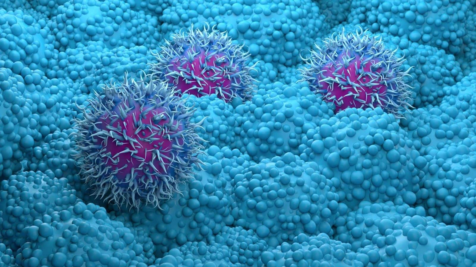
New imaging technique offers insights into how cancer cells work
What's the story
Scientists from the University of Surrey, in collaboration with University College London, GSK, Yokogawa, and Sciex, have developed an innovative imaging method for cancer. This groundbreaking technique provides a detailed view of the lipid content within individual cancer cells. The research aims to explore the significant role of lipids in cancer cell growth and proliferation as stated by Melanie Bailey, a senior author at the University of Surrey.
Technological breakthrough
How the researchers isolated and marked the cells
The research team utilized the Single Cellome System SS2000 from Yokogawa to isolate intact pancreatic cancer cells from a sample. These cells were then marked with a fluorescent dye that highlighted their lipid content. In collaboration with Sciex, a leading mass spectrometry manufacturer, they then developed a unique mass spectrometry technique. This method allowed them to dissect the lipids and reveal their exact composition, Bailey explained.
Research findings
Lipid profiles could help understand how cancer cells react
The team's research, published in the Analytical Chemistry journal, revealed significant variations in lipid profiles among different cancer cells. They also observed how these lipids adapt to environmental changes. This discovery could lead to significant advancements in cancer research and treatment strategies. The innovative imaging technique also holds potential for research beyond cancer, particularly in fields where understanding lipid behavior in individual cells is crucial.
Information
The technique is being explored for diverse fields
Bailey's team is already collaborating with other researchers to study lipids in single cells across diverse areas such as immunity, infectious diseases, and circadian rhythm studies.