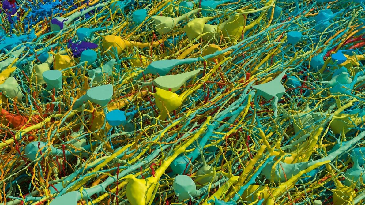
Google, Harvard create the most detailed map of human brain
What's the story
In a groundbreaking collaboration, Harvard biologists and Google have created the most comprehensive map of human brain connections ever seen. The project, which spanned a decade, utilized a cubic millimeter of the human cerebral cortex at the subcellular level. This remarkable achievement is expected to revolutionize future brain science research. The sample used for this study was obtained from a 45-year-old woman undergoing epilepsy surgery in 2014.
Project details
Map includes approximately 57,000 cells and 150 million synapses
The team of biologists and machine-learning (ML) experts spent a decade creating an interactive map of the brain tissue. This intricate diagram includes approximately 57,000 cells and 150 million synapses. The map reveals cells that wrap around themselves, pairs of cells that appear mirrored, and egg-shaped "objects" that defy categorization. Daniel Berger, a lead researcher on the project, believes this high-resolution mapping could help identify "rules of wiring" between neurons.
Methodology
Innovative techniques used in brain mapping
The brain map was created by taking subcellular pictures of the tissue using electron microscopy. The tissue from the woman's brain was stained with heavy metals that attach to lipid membranes in cells, making them visible under an electron microscope. It was then embedded in resin and cut into thin slices, just 34 nanometers thick. This process transformed a 3D problem into a 2D one for easier mapping, according to Berger.
Tech contribution
Google's machine learning algorithms aid in brain mapping
After obtaining the images, the Harvard researchers collaborated with a team at Google led by Viren Jain. The tech giant's team used machine-learning algorithms to align the 2D images and produce 3D reconstructions with automatic segmentation. This process differentiated and categorized components within an image, such as different cell types. Some of this segmentation required manual redrawing of some tissue by hand to further inform the algorithms, a task undertaken by Berger who worked closely with Google's team.
Tech advancement
Digital technology enhances visibility
Berger used digital technology to color the cells in the tissue sample differently based on their size. This method is a significant improvement over conventional methods of imaging neurons, which often leave some elements of nervous tissue hidden. The smallest cells were colored blue and the biggest ones red, with all other cells falling on a color spectrum. This technique helped researchers identify the brain's six cortical layers and white matter.
Research hurdles
Challenges faced during the project
One of the challenges faced by the researchers was proofreading the automatic segmentation. This involved manually checking every part of the 3D map for segmentation errors, a task that proved to be a significant challenge due to the large datasets generated. Despite these challenges, Berger noted that certain cells seemed "really interested in contacting." The team also identified abnormal cells during their research, further highlighting the complexity of this project.