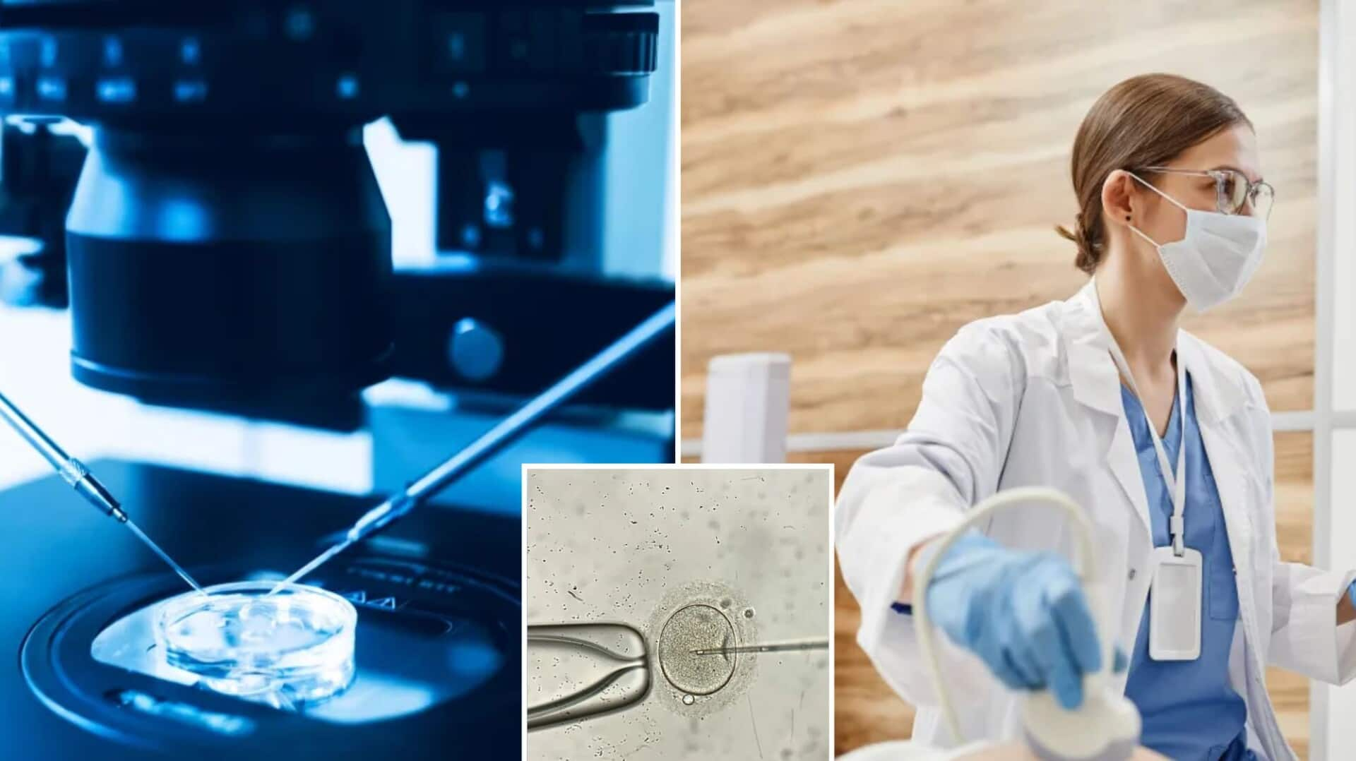
Breakthrough for IVF? New 3D imaging method could boost outcomes
What's the story
A team of researchers, led by Dr. Bo Huang from the Reproductive Medicine Center of Tongji Hospital in China, has developed a 3D imaging model for early-stage embryos.
This innovative technology could potentially simplify the conception process for women undergoing in vitro fertilization (IVF).
The study focused on blastocysts—embryos that are 5 or 6 days old—and revealed previously unknown cell features associated with successful pregnancies.
Key indicators
3D model sheds light on blastocyst features for IVF success
Huang's study reveals that the 3D shape of a blastocyst's inner cell mass, its position, and the arrangement of surrounding cells can be crucial indicators of successful pregnancies.
This information was previously unknown.
However, determining which embryos have the highest likelihood of resulting in a successful birth remains challenging.
In 2022, approximately 92,000 births in the US were from IVF, accounting for about 2.5% of all births.
IVF challenges
Study highlights limitations of current IVF procedures
During IVF, eggs are collected from a woman and fertilized with sperm in a lab to create embryos.
These embryos are then transferred into the uterus.
Despite genetic testing of embryos before transfer, the success rate for genetically healthy embryos is only between 60% to 65%.
This percentage decreases further if the woman is older or has uterine conditions that make it difficult for an embryo to implant.
Research findings
3D imaging model proves highly accurate in study
The study involved women under 40 with a thick uterine lining and no more than one failed embryo transfer.
Using EmbryoScope+, a technology that monitors embryo development, researchers took detailed images of 2,141 blastocysts.
Huang's team found their model to be 90% accurate when compared with fluorescence imaging of the blastocysts.
This new method offers a significant improvement over traditional 2D methods that lack depth and comprehensive indicators.
Clinical application
New 3D model bridges gap in embryo evaluation methods
Huang explained that while some 3D methods exist, they are not practical or safe for clinical use.
The new study introduces a clinically applicable 3D evaluation method and uncovers previously unrecognized features of blastocysts.
The research was presented at the annual meeting of the European Society of Human Reproduction and Embryology (ESHRE) in Amsterdam and published in the journal Human Reproduction.