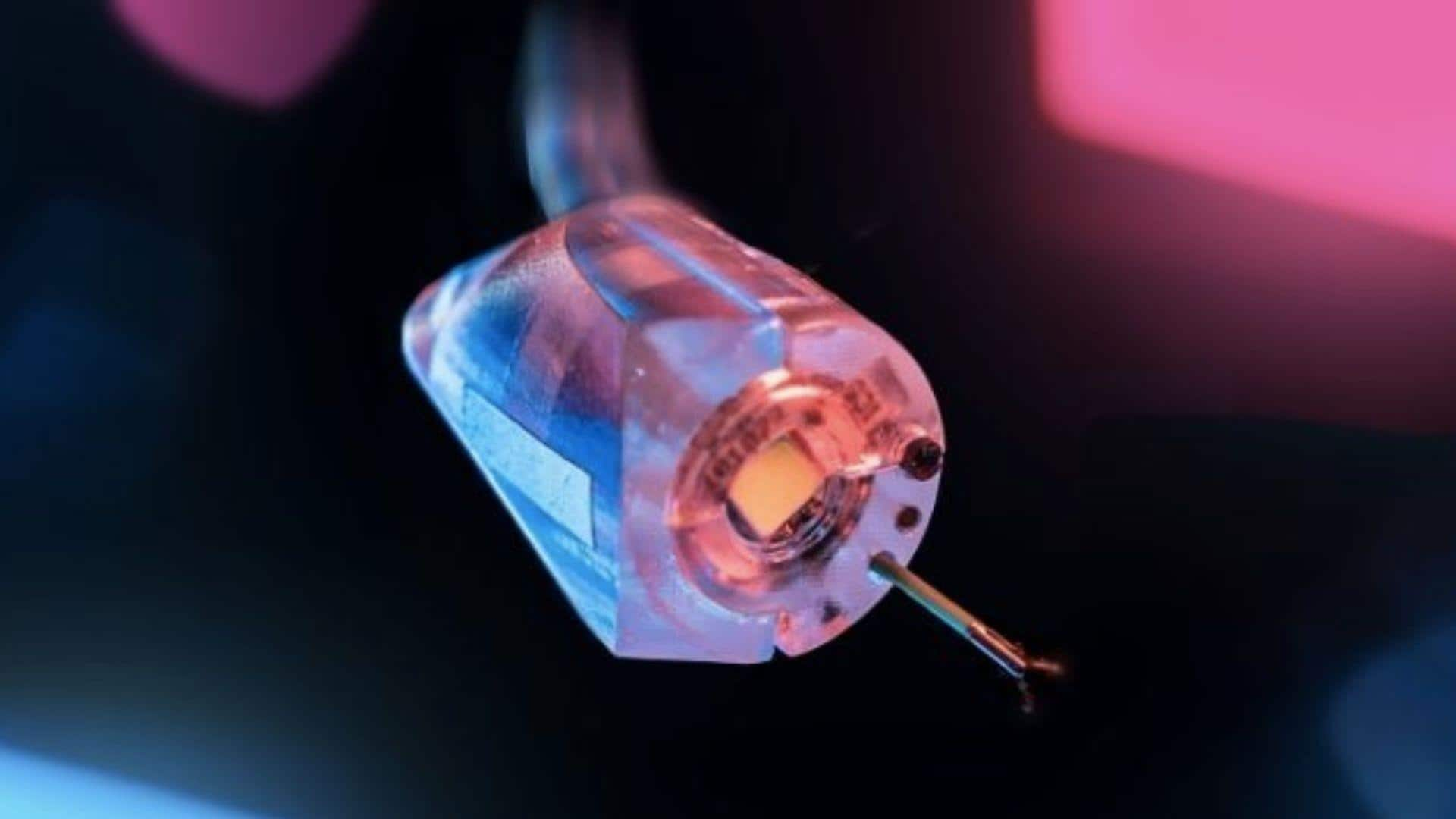
Magnetic robot helps perform 'virtual biopsies' for early cancer detection
What's the story
A team of engineers at the University of Leeds has developed a groundbreaking magnetic robot that could revolutionize early cancer detection.
The tiny machine can produce high-resolution, three-dimensional ultrasound images from deep inside the body, including regions like the gastrointestinal tract or gut.
This innovation allows 'virtual biopsies,' non-invasive scans that give immediate diagnostic data to doctors.
Virtual biopsies
New approach to cancer diagnosis
With its unique design, the robot enables doctors to detect, stage, and even treat lesions in one go.
This means no more physical biopsies and a potential revolution in the diagnosis and treatment of different kinds of cancer.
The researchers incorporated an oloid shape into a new magnetic flexible endoscope (MFE), which had a small high-frequency imaging device for detailed 3D internal tissue images.
Innovation
Breakthrough in medical imaging
Professor Pietro Valdastri, who led the research, said this study allows reconstructing a 3D ultrasound image from a probe deep inside the gut, something that has never been done before.
The robot's design is inspired by embodied intelligence and the unique geometry of developable rollers.
The oloid shape with its axial asymmetry and sinusoidal motion facilitates rolling when precisely controlled by an external magnetic field.
Study validation
A step forward in non-invasive imaging
The study, published in Science Robotics, details a versatile closed-loop control model to ensure precise magnetic manipulation of an oloid-shaped robot.
This capability was validated by integrating a 28MHz micro-ultrasound array into endoluminal applications for virtual biopsies and non-invasive real-time histological imaging.
The imaging device creates high-resolution 3D reconstructions of scanned areas, enabling clinicians to create cross-sectional images like those produced by standard biopsies.
Patient experience
Enhancing patient comfort and reducing anxiety
While 3D ultrasound imaging is already employed in blood vessels and rectal examinations, this study paves way for deeper scans in the GI tract.
Nikita Greenidge from Leeds's STORM Lab emphasized that this innovation enables doctors to diagnose and treat in one procedure, removing the wait between diagnosis and intervention.
This not only enhances patient comfort but also reduces waiting times, repeat procedures, and anxiety of waiting for potential cancer results.