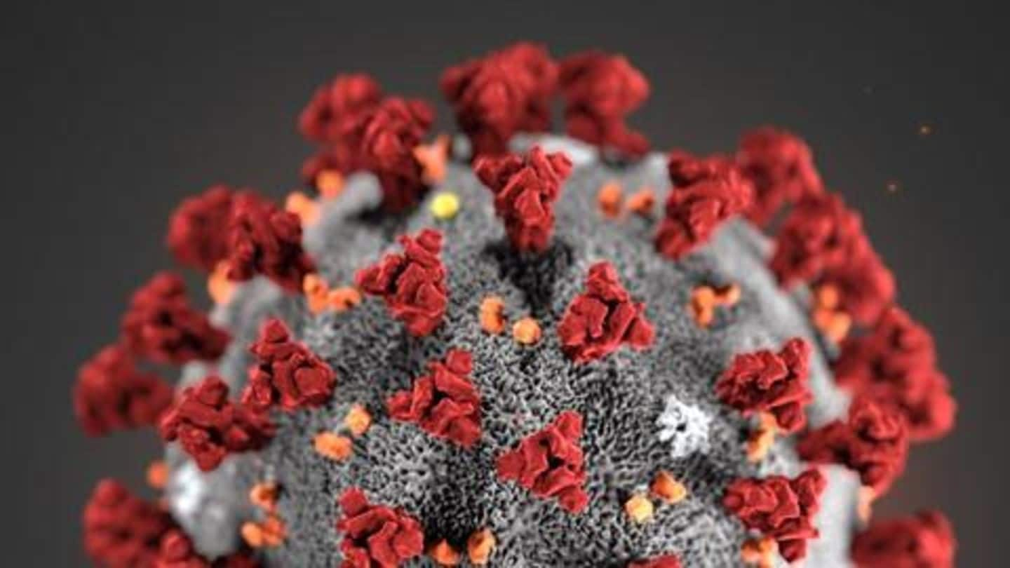
In a first, Indian scientists capture image of novel coronavirus
What's the story
Scientists in Pune have managed to capture the image of the novel coronavirus responsible for the current COVID-19 outbreak. This is the first time that Indian scientists have managed to capture an image of the pathogen, called SARS-CoV-2. For the image, the scientists used the virus isolated from India's first coronavirus patient, a Kerala woman who tested positive on January 30.
Details
Image published in Indian Journal of Medical Research
The image of SARS-CoV-2 was captured using a transmission electron microscope (TEM) and has been published in the latest edition of the Indian Journal of Medical Research (IJMR). The sample was isolated from the Kerala woman, who studying in China's Wuhan—the epicenter of the outbreak. Reportedly, gene sequencing of the sample had earlier found a 99.98% match with the virus in Wuhan.
Methodology
Here's how the scientists captured the microscopic image
A 500μl sample confirmed positive for SARS-CoV-2 nucleic acid by real-time polymerase chain reaction (PCR) was centrifuged to remove debris. The supernatant (liquid overlying precipitated solids after centrifugation) was removed, fixed using 1% glutaraldehyde, and adsorbed onto a carbon-coated 200 mesh copper grid. Sodium phosphotungstic acid was used for negative staining. It was then examined under 100 kV accelerating voltage in a TEM.
Observation
One virus particle had 'very typical' coronavirus features
Seven negative-stained virus particles were found with morpho-diagnostic features of a coronavirus-like particle. One well-preserved 75nm virus particle showed "very typical" coronavirus features. It had distinct envelope projection ending in round 'peplomeric' structures. A peplomer is glycoprotein protrusion in the outer protein coat of a virus. These peplomers give the coronaviruses a crown-like appearance, hence the name 'coronavirus' (corona is Latin for crown).
Applications
Image could help further studies in virus origins, mutations, etc.
Indian Council of Medical Research (ICMR) former Director-General Dr. Nirmal K Ganguly told Hindustan Times, "These images are critical to study mutations in clinical samples and help identify the genetic origin and evolution of the virus." Ganguly said it could help answer how the virus jumped from animals to humans, how human-to-human transmission began, if the virus is still mutating, and other questions.